Retinal Tears

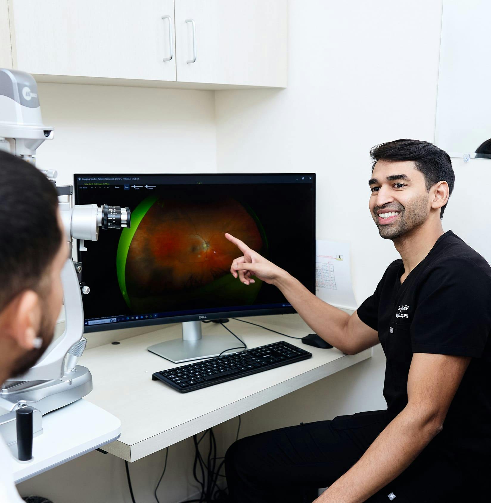
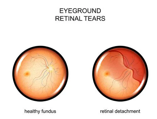
Our ophthalmologist  is available for retinal tear treatment.

Retinal Tear Diagram

Understanding Retinal Tears

Your retina is made up of photosensitive cells that communicate with your brain, thus allowing you to see. It's quite thin and sometimes tears are part of the aging process. A tear will allow fluid to enter the inside of the eye, leading to a retinal detachment. That's why It's vital to have the problem addressed quickly by our retinal surgeon, Dr. Vinod Jindal.

How do Retinal Tears Happen?

The eye is filled with vitreous, a gel-like substance that breaks down and becomes more watery as we age. In people older than 60 years of age, this process often causes the vitreous to separate from the back of the eye. When this occurs, you are at risk of developing a retinal tear. The vitreous moves more easily when detached from the back of the eye, so it's very easy to develop a tear in your retina during the course of common, everyday movements. Some people are at higher risk for retinal tears, including those with:

  • Nearsightedness,
  • Retinal thinning,
  • Previous cataract surgery,
  • A history of eye trauma,
  • A family history of retinal tears,
  • Retinal tears in the other eye.

Symptoms of a Retinal Tear

Symptoms of retinal tears include flashes of light in the eye and seeing floaters in your eye. Retinal tears can also cause bleeding into the eye, which causes multiple floaters and vision loss.

Retinal Tear Treatment

Our doctors can diagnose a retinal tear during an eye exam.  If a retinal tear is present, immediate treatment is required to prevent retinal detachment.  Retinal tears are common, but they should be treated quickly to avoid vision loss. 

Retinal tears are corrected through the creation of scar tissue around the tear to act in as an adhesive. Scar tissue prevents fluid from entering the eye and prevents the tear from progressing. There are two methods to build scar tissue. 

  • First and most common, laser photocoagulation creates tiny burns around the tear. Scar tissue is formed as the burns heal. 
  • The less common method is cryopexy.  This may be recommended if the presence of blood prevents our doctors from seeing the tear. A cryoprobe is placed on the outside of the eye and becomes very cold, creating freeze burn around the tear. Scar tissue is created during the healing process. 

Following either type of treatment, you will receive instructions from Dr. Jindal to optimize best healing.  It is important that you follow these guidelines to yield the best visual outcome possible.

Contact Us

We look forward to hearing from you.

Locations

Find us on the map

Hours of Operation

Our Regular Schedule

Main Office

Monday:

8:00 am-5:00 pm

Tuesday:

8:00 am-5:00 pm

Wednesday:

8:00 am-5:00 pm

Thursday:

8:00 am-5:00 pm

Friday:

8:00 am-5:00 pm

Saturday:

Closed

Sunday:

Closed